Many people are aware of the concept that their blood group influences their interaction with the environment: intestinal secretions of blood group antigens affect a person’s interaction with foods through being a marker of self recognised by the immune system. Similar to the way that a transfusion of blood from the wrong blood group causes a powerful IgM immune reaction, food lectins can also lead to a reaction (albeit less immunologically strong) by stimulating release of IL4 and IL13 from basophils, potentially leading to type I allergy. [1] Depending on diet, this could result from lectins incompatible with specific blood group glycoproteins, which would create an immune response individual to the person’s blood group: lectins from some foods are used to preferentially agglutinate specific glycoprotein antigens when testing saliva. [2]
Secretor status is under genetic control: 15-20% of people of Western European descent are unable to secrete their blood group antigens due to a homozygous mutation on the fucosyltransferase 2 (FUT2) gene at the rs601338 SNP. The FUT2 (secretor) gene is expressed predominantly in secretory tissues, giving rise to glycoprotein products in mucin secretions. [3] This mutation is rare in Chinese and Japanese populations, and instead the more common homozygous FUT2 rs1047781 missense mutation is responsible for dramatically decreased expression of ABH antigens (partial ABH secretors). [4]
Non-secretor status is associated with immunity to norovirus, [5] higher vitamin B12 levels, [PMID: 18776911] and secretor status also affects phagocytic activity of the leukocytes in a manner that places non-secretors at an advantage from increased activity, [6] in addition to the influence of the ABO blood group on phagocytosis. [7] Many disease risks are also associated with non-secretor status: antigen barrier function favours secretors, the free ABH antigen on the mucosal barriers of ABH secretors acts as an effective anti-adhesive mechanism against ABH specific bacterial fimbriae lectins. [8] Non-secretors may have an increased risk for Crohn’s disease, [9] type 1 diabetes, [10] and vaginal candidiasis. [11]
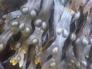
One way to determine whether a person is a secretor of their blood group is to test their saliva for their ABO blood group antigens. This method is not commonly available, as a supply of red blood cells is needed for testing the saliva. Another way to find secretor status is to test for Lewis blood group. The Lewis a (Le a) antigen is normally secreted into the blood and then adsorbed onto red blood cells, [12] where it can be agglutinated with anti-Lewis a reagent. Fucose is a common sugar, found in seaweeds such as fucus vesiculosus (bladderwrack), and also forms the terminal sugar of the H antigen in people with blood group O. Fucosyltransferase enzymes can attach the fucose molecule onto another sugar or glycoprotein through fucosylation, such as that of the blood group A or B antigen or Lewis antigens. If there is a working copy of the FUT2 SNP the Lewis a antigen will be catalysed into Lewis b by the FUT2 enzyme. Consequently those with a functional FUT2 enzyme don’t have any Lewis a antigen on their erythrocytes, but they will have the Lewis b antigen, and therefore a finding of Lewis b on erythrocytes indicates an ABH secretor. The problem arises with the small number of people who don’t have the Lewis blood group antigens on their blood cells. This is similar to the rare ‘Bombay’ blood group, which results in a loss of production of ABH antigens on erythrocytes (from loss of FUT1 function): the fucosyltransferase 1 (FUT1) gene is expressed predominantly in erythroid tissues, giving rise to FUT1 (H enzyme) giving rise to products found on erythrocytes. About 5% of the European population (and more in some other populations) lack a functional fucosyltransferase 3 (FUT3) enzyme. These FUT3 negative people are unable to make any Lewis antigens. They may be either ABH secretors or non-secretors, but the Lewis test cannot be used to determine secretor status due to the lack of any Lewis antigen for agglutination.
People with no Lewis antigens are classed as Lewis negative, however this phenotype might not always be only caused by FUT3 mutation. Erythrocyte membranes have been found to lose their Lewis antigens during pregnancy and during diseases such as cancer: individuals have been identified who change from Lewis positive to Lewis negative on erythrocytes, although they persistently express Lewis enzyme activity in saliva. The reason for this change has been attributed to an increased level of circulating lipoproteins during the burden of disease or pregnancy, which alter the balance between production of Lewis glycolipids, transport in lipoproteins, and incorporation into erythrocyte membranes. Fucosyltransferase activity in saliva is variable, being lower in FUT3 heterozygotes than it is in homozygous wild-type individuals, and those with mutation in the FUT2 gene (non-secretors) do not fucosylate the Lewis a structure to H and the Lewis b, in competition with a sialyltransferase. [13]
There are also epigenetic influences on FUT3 expression. Certain cancer markers are not found in patients with FUT3 mutation: it is not thought useful to measure the CA19-9 titer of Lewis negative cancer patients. A study found that Lewis-negative individuals consisting of a homozygous negative FUT3 genotype had completely negative CA19-9 values, irrespective of the FUT2 secretor genotype. [14] Very few Lewis-positive patients exhibit positive DU-PAN-2 values. [15]
Although Lewis negative status may be protective against Rotavirus, [16] it is also linked with markers of inflammation: WBC, hs-CRP and ESR were significantly elevated, and rheological parameters (RBC aggregation, plasma viscosity) were found to be abnormal in Lewis negative subjects. [17] Lewis negative men were found to have a higher systolic blood pressure (6 mm Hg), higher values for BMI (8%) and total body fat mass than Lewis positive individuals. [18] Lewis negative status is a genetic risk factor for ischemic heart disease (IHD), particularly in men, and is associated with high triglycerides. Lewis negative status also confers protection from IHD with moderate alcohol intake: Studies found that the risk of IHD was negatively correlated with alcohol consumption. [19] The authors suggest that alcohol consumption may modify insulin resistance in Le(a-b-) men. [20] Asthma is related to both non-secretor and Lewis negative phenotypes, and low lung function values have been observed in Lewis negative non-secretors. Alcohol intake is also protective against asthma in Lewis negative individuals, [21] but Lewis negative individuals are more likely to suffer from alcoholism. [22] Lewis negative phenotype confers a three times greater risk of diabetes, [23] and an increased risk for Sjögren’s syndrome. [24] The intestinal microbiota of individuals with Lewis negative blood groups were reported to contain a less rich and diverse range of bacteria than those with Lewis a phenotype. [25] Urinary tract infections in women are more common amongst non-secretors, and most common in Lewis negative individuals. [26] Polymorphisms in FUT3 and its intestinal expression might be associated with pathogenesis of ulcerative colitis. [27]
Despite the differences in disease risk between ABH secretors and non-secretors, clinical experience suggests that Lewis negative individuals appear to have unique interactions with certain disease states. [8] Opus 23 Pro [28] provides algorithms in the Lumen app to find secretor status from the FUT2 rs601338 SNP, and to estimate Lewis status from 23andMe raw data based on the SNPs available for FUT3 (all except one of the most common FUT3 SNPs resulting in Lewis negative status are typically reported by 23andMe). This can give the practitioner another level of insight into the likely glycosylation levels, immune status, inflammatory and health risks of the patient, as well as the likelihood of relevance of testing for tumour markers.
- Haas H, Falcone FH, Schramm G, et.al. Dietary lectins can induce in vitro release of IL-4 and IL-13 from human basophils. Eur J Immunol. 1999 Mar;29(3):918-27. PMID: 10092096.
- Albertolle ME, Hassis ME, Ng CJ, et. al. Mass spectrometry-based analyses showing the effects of secretor and blood group status on salivary N-glycosylation. Clin Proteomics. 2015 Dec 30;12:29. doi: 10.1186/s12014-015-9100-y. PMID: 26719750; PMCID: PMC4696288.
- Prakobphol A, Leffler H, Fisher SJ. The high-molecular-weight human mucin is the primary salivary carrier of ABH, Le(a), and Le(b) blood group antigens. Crit Rev Oral Biol Med. 1993;4(3-4):325-33. PMID: 7690601.
- Hu D, Zhang D, Zheng S, et. al. Association of Ulcerative Colitis with FUT2 and FUT3 Polymorphisms in Patients from Southeast China. PLoS One. 2016 Jan 14;11(1):e0146557. doi: 10.1371/journal.pone.0146557. PMID: 26766790; PMCID: PMC4713070.
- Lindesmith L, Moe C, Marionneau S, et. al. Human susceptibility and resistance to Norwalk virus infection. Nat Med. 2003 May;9(5):548-53. PMID: 12692541.
- Tandon OP, Bhatia S, Tripathi RL, Sharma KN. Phagocytic response of leucocytes in secretors and non-secretors of ABH (O) blood group substances. Indian J Physiol Pharmacol. 1979 Oct-Dec;23(4):321-4. PMID: 528036.
- Tandon OP. Leucocyte phagocytic response in relation to abo blood groups. Indian J Physiol Pharmacol. 1977 Jul-Sep;21(3):191-4. PMID: 612601.
- D’Adamo PJ, Kelly GS. Metabolic and immunologic consequences of ABH secretor and Lewis subtype status. Altern Med Rev. 2001 Aug;6(4):390-405. Review. PMID: 11578255.
- McGovern DP, Jones MR, Taylor KD, et. al. International IBD Genetics Consortium. Fucosyltransferase 2 (FUT2) non-secretor status is associated with Crohn’s disease. Hum Mol Genet. 2010 Sep 1;19(17):3468-76. doi: 10.1093/hmg/ddq248. PMID: 20570966; PMCID: PMC2916706.
- Smyth DJ, Cooper JD, Howson JM, et. al. FUT2 nonsecretor status links type 1 diabetes susceptibility and resistance to infection. Diabetes. 2011 Nov;60(11):3081-4. doi: 10.2337/db11-0638. PMID: 22025780; PMCID: PMC3198057.
- Hurd EA, Domino SE. Increased susceptibility of secretor factor gene Fut2-null mice to experimental vaginal candidiasis. Infect Immun. 2004 Jul;72(7):4279-81. PMID: 15213174; PMCID: PMC427463.
- Henry S, Oriol R, Samuelsson B. Lewis histo-blood group system and associated secretory phenotypes. Vox Sang. 1995;69(3):166-82. Review. PMID: 8578728.
- Orntoft TF, Vestergaard EM, Holmes E, et. al. Influence of Lewis alpha1-3/4-L-fucosyltransferase (FUT3) gene mutations on enzyme activity, erythrocyte phenotyping, and circulating tumor marker sialyl-Lewis a levels. J Biol Chem. 1996 Dec 13;271(50):32260-8. PMID: 8943285.
- Narimatsu H, Iwasaki H, Nakayama F, et. al. Lewis and secretor gene dosages affect CA19-9 and DU-PAN-2 serum levels in normal individuals and colorectal cancer patients. Cancer Res. 1998 Feb 1;58(3):512-8. PMID: 9458099.
- Vestergaard EM, Hein HO, Meyer H, et.al. Reference values and biological variation for tumor marker CA 19-9 in serum for different Lewis and secretor genotypes and evaluation of secretor and Lewis genotyping in a Caucasian population. Clin Chem. 1999 Jan;45(1):54-61. PMID: 9895338.
- Nordgren J, Sharma S, Bucardo F, et. al. Both Lewis and secretor status mediate susceptibility to rotavirus infections in a rotavirus genotype-dependent manner. Clin Infect Dis. 2014 Dec 1;59(11):1567-73. doi: 10.1093/cid/ciu633. PMID: 25097083; PMCID: PMC4650770.
- Alexy T, Pais E, Wenby RB, et al. Abnormal blood rheology and chronic low grade inflammation: possible risk factors for accelerated atherosclerosis and coronary artery disease in Lewis negative subjects. Atherosclerosis. 2015;239(1):248-251. doi:10.1016/j.atherosclerosis.2015.01.015.PMID: 25626016; PMCID: PMC4331217
- Clausen JO, Hein HO, Suadicani P, et. al. Lewis phenotypes and the insulin resistance syndrome in young healthy white men and women. Am J Hypertens. 1995 Nov;8(11):1060-6. PMID: 8554728.
- Hein HO, Sørensen H, Suadicani P, Gyntelberg F. Alcohol intake, Lewis phenotypes and risk of ischemic heart disease. The Copenhagen Male Study. Ugeskr Laeger. 1994 Feb 28;156(9):1297-302. PMID: 8009753.
- Hein HO, Sørensen H, Suadicani P, Gyntelberg F. Alcohol consumption, Lewis phenotypes, and risk of ischaemic heart disease. Lancet. 1993 Feb 13;341(8842):392-6. PMID: 8094167.
- Kauffmann F, Frette C, Pham QT, Nafissi S, Bertrand JP, Oriol R. Associations of blood group-related antigens to FEV1, wheezing, and asthma. Am J Respir Crit Care Med. 1996 Jan;153(1):76-82. PMID: 8542166.
- Cruz-Coke R. Genetics and alcoholism. Neurobehav Toxicol Teratol. 1983 Mar-Apr;5(2):179-80. PMID: 6346123.
- Melis C, Mercier P, Vague P, Vialettes B. Lewis antigen and diabetes. Rev Fr Transfus Immunohematol. 1978 Sep;21(4):965-71. PMID: 734307.
- Manthorpe R, Staub Nielsen L, Hagen Petersen S, Prause JU. Lewis blood type frequency in patients with primary Sjögren’s syndrome. A prospective study including analyses for A1A2BO, Secretor, MNSs, P, Duffy, Kell, Lutheran and rhesus blood groups. Scand J Rheumatol. 1985;14(2):159-62. PMID: 4001887
- Wacklin P, Tuimala J, Nikkilä J. Faecal microbiota composition in adults is associated with the FUT2 gene determining the secretor status. PLoS One. 2014 Apr 14;9(4):e94863. doi: 10.1371/journal.pone.0094863. PMID: 24733310; PMCID: PMC3986271.
- Sheinfeld J, Schaeffer AJ, Cordon-Cardo C, Rogatko A, Fair WR. Association of the Lewis blood-group phenotype with recurrent urinary tract infections in women. N Engl J Med. 1989 Mar 23;320(12):773-7. PMID: 2922027.
- Hu D, Zhang D, Zheng S, et. al. Association of Ulcerative Colitis with FUT2 and FUT3 Polymorphisms in Patients from Southeast China. PLoS One. 2016 Jan 14;11(1):e0146557. doi: 10.1371/journal.pone.0146557. PMID: 26766790; PMCID: PMC4713070.
- Opus 23 Pro genetic analysis and reporting software by Dr P. D’Adamo www.opus23.com.
